



Featured
oi-AK Khan

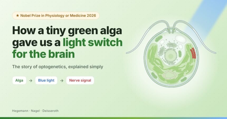
A simple question about pond algae led scientists to a way of turning brain cells on and off with a flash of blue light. This is the story of optogenetics, told step by step.
How does a one-celled alga “see” light so fast?
A protein that opens like a gate when light hits it.
Put that gate into nerve cells and control them with light.
A scientist asks: how does an alga see?
In the early 1990s, Peter Hegemann, a researcher in Germany, became fascinated by Chlamydomonas, a green alga so small that about 65 of them lined up would only span one millimetre. It has no brain and no eyes, yet it can do something clever: it swims towards light.
Stir these algae into a dish of water and the water turns faintly green. Shine a lamp on one side, and the green colour slowly drifts towards the light, because millions of algae are swimming that way.

What is the eye spot? A small orange-red patch on the cell. It contains retinal, the same kind of light-catching molecule found in our own eyes. Think of it as the alga’s single, very simple “pixel”.
What are flagella? Two thin, whip-like hairs on the front of the alga that it beats like swimming arms to move and steer through water.
The alga reacts faster than our own eyes
Hegemann used extremely thin electrodes to listen in on the tiny electrical signals inside the alga. He found that just half a millisecond after light hit the eye spot, an electrical pulse appeared.

Every cell has tiny gates in its outer wall
Every living cell is wrapped in a thin, oily skin called the cell membrane. Nothing charged can slip through it on its own. Instead, the membrane holds tiny protein doors called ion channels. When a door opens, charged particles flow from outside the cell to inside the cell.
⊕⊕⊕⊕
→
The gate (ion channel)A protein door in the cell wall. Closed or open.
→
⊕ ⚡
Called ions: atoms that carry a tiny electric charge, like sodium (from salt), potassium and calcium dissolved in the water in and around our cells.
At rest, the inside of a cell is slightly negative and has fewer of these ions. Positive ions are pulled towards it, like water rushing downhill the moment a dam gate opens.
That sudden rush flips the cell’s charge, making a small electric signal. In nerve cells, this is how messages travel. In the alga, it tells the flagella how to beat so it steers towards light.
How fast is fast? A speed race
ms = millisecond, one thousandth of a second. Bars are drawn to scale.
Human eye
Light → molecule A → B → C → … → gate opens
Like a long relay race
⇄
Alga (Hegemann’s idea)
Light → gate opens
One runner does everything
In our eyes, light sets off a chain reaction of many molecules before the gate finally opens and an electric signal travels to the brain, so that we see. Hegemann reasoned that the alga must skip all those steps. His bold idea: a single protein both catches the light and is the gate itself.
Why people doubted him Scientists knew many ion channels, gates in the cell wall that let charged particles pass from outside the cell to inside. But none of them could be opened by light on its own. It took about ten years to prove him right.
Finding the light-operated gate
The protein was fragile and fell apart when taken out of the alga. The breakthrough came around the year 2000, when Japanese researchers published the alga’s genetic code. Hegemann’s team spotted two genes that looked like known light-catching proteins.
He sent them to Georg Nagel, a colleague in Frankfurt who studied proteins using frog eggs. Nagel injected each gene into frog eggs, which then built the proteins and placed them on their surface. When he shone light on the eggs, the proteins opened, exactly as Hegemann had predicted a decade earlier.

They named the proteins channelrhodopsin-1 and channelrhodopsin-2 (“channel” = gate, “rhodopsin” = light-sensing protein). The second one turned out to be the star.
The simple science, in four steps
1
Wall
Every cell is wrapped in a thin oily skin called a membrane.
2
Gate
Channelrhodopsin sits in this wall like a closed door.
3
Light
Blue light hits it and the door swings open in 0.2 ms.
4
Spark
Positive ions flow in, creating a tiny electric pulse.
The key insight (2003) They put the gene into kidney cells from humans and from hamsters (small rodents, close cousins of rats and mice). Those cells also became light-sensitive. The gate works in any cell it is placed in. Hegemann and Nagel suggested it could become a tool to switch cells on with light.
A doctor turns it into a switch for nerve cells
Karl Deisseroth trained as a doctor at Stanford University. Working in a psychiatric clinic, he met patients with depression, autism and schizophrenia, and saw how little the available treatments helped. He wanted to understand how specific brain cells cause feelings and behaviour, in a living brain.
The brain works through electrical signals in nerve cells. If he could put the light-gate into chosen nerve cells, a flash of light would make exactly those cells fire, and nothing else.
Step one: rat nerve cells in a dish
Deisseroth wrote to Georg Nagel and asked for the DNA (the genetic instructions) for channelrhodopsin-2. He then grew nerve cells taken from rats in petri dishes and added the gene to them.
He was worried. Nerve cells are delicate, and a gene from pond algae might harm them. It did not: the rat nerve cells simply read the new instructions and built the light-gate into their own walls, as if it were one of their own proteins.
1
Add the gene
Alga DNA is put into rat nerve cells growing in a dish.
2
Cells build gates
The cells make channelrhodopsin-2 and place it in their walls.
3
Flash blue light
The gates open and positive ions rush in.
4
Nerve fires
A real nerve signal starts and passes on to other nerve cells.

Think of it like this The brain is a city with 90 billion light bulbs, all wired together. Old methods could only switch off whole neighbourhoods. Optogenetics lets scientists install a special switch in one chosen type of bulb and flick only those on with a torch.
Step two: a living, moving mouse
A dish is not a brain. The real test was whether light could control nerve cells inside a living animal. Deisseroth chose the motor cortex, the part of the brain that sends “move!” commands to the muscles of the body.
The scientists chose which cells would get the light-gate. They packed the alga gene inside a harmless virus, used only as a delivery vehicle, and injected it into the motor cortex of mice.
Those chosen cells then followed the new instructions and built the light-gates themselves, just as the rat cells in the dish had. The other cells nearby ignored the gene, so they had no light-gates and did not respond to light. To get light deep inside the brain, they passed a hair-thin optical fibre (a tiny glass thread that carries light) through a small hole in the skull.
What happened when the light came on? The lit-up motor cells fired, sending their “move!” command down to the face, and the mouse’s whiskers moved. When the light went off, the movement stopped. The mouse had not decided to move its whiskers; the scientists’ light had given the order.
What are whiskers? The long, stiff hairs on a mouse’s snout. Mice sweep them back and forth to feel their way around, much like we use our fingertips in the dark.
For the first time, a scientist could say: these exact cells cause this exact movement, and prove it with the flick of a light switch.
Working with other researchers, Deisseroth put the light-gate into a recently discovered type of nerve cell (now known as hypocretin or orexin cells) that was suspected to control wakefulness.
When the mice were asleep and light was shone on these cells, the mice woke up. The light confirmed what scientists had only guessed: these cells act like the brain’s alarm clock.
Switching on a single memory
Every memory is stored in a particular group of nerve cells that are active together when the memory forms. Scientists call this group an engram, a kind of memory “footprint” in the brain. Together with Susumu Tonegawa (a Nobel laureate himself, in 1987), Deisseroth set out to find one.
Making the memory. A mouse was placed in a box where it had an unpleasant, frightening experience. While the fear memory was forming, the scientists noted which nerve cells in the memory area of the brain were active, and used a genetic trick to give exactly those cells the light-gate.
Waking the memory. Later, the mouse was moved to a completely different, safe box with nothing to be afraid of. Then the light was switched on, activating only the tagged memory cells.
The result. The mouse suddenly froze, the way mice naturally do when they are afraid, even though there was no danger at all. The light had made it relive the frightening memory.
Why this was a landmark It was the first experiment to show exactly which nerve cells are needed for one specific memory. A memory was no longer a vague idea; it could be found, tagged and switched on.
How it grew, step by step
Early 1990s
Hegemann measures the alga’s ultrafast light response and proposes the one-protein gate idea.
2003
Hegemann and Nagel describe channelrhodopsin-2 and show it works in other cells.
2005
Deisseroth makes rat nerve cells in a dish fire with blue light.
2006
The method gets its name: optogenetics (opto = light, genetics = genes).
2007
It works in living mice: whiskers move on command, and sleeping mice are woken with light.
2012
With Susumu Tonegawa, cells holding a single fear memory are switched on, and the mice act afraid with no danger present.
Who did what
The question
Peter Hegemann
Asked why the alga reacts so fast and predicted a single light-operated gate.
The proof
Georg Nagel
Used frog eggs to show the alga’s proteins really are gates that open with light.
The tool
Karl Deisseroth
Put the gate into nerve cells and made it work in living brains.
A new map of the brain
For a century, brain maps were rough sketches. Scientists could see that an area was active, but not prove it caused a behaviour. Worse, cells doing completely different jobs sit tangled together like wires in a bundle. With optogenetics, scientists can light up one cell type in that bundle and watch what the animal does. That is cause and effect. Here is some of what they found.
◉
Senses & needs: thirst, hunger, pain
Scientists found small groups of cells that work like a body’s “need” buttons. Switch on certain thirst cells and a mouse that has just had plenty to drink starts drinking eagerly again, as if parched.
Similar circuits have been mapped for eating, pain and attention. Why it matters: it shows that a feeling as basic as “I’m thirsty” is produced by specific, findable cells.
★
Reward & social life
When light activates the brain’s reward cells while a mouse is in one corner of its box, the mouse keeps going back to that corner. It has learned that the spot “feels good”.
Circuits behind social behaviour, such as interest in other mice, have also been traced. Why it matters: reward circuits are at the heart of motivation, habits and addiction.
♡
Feelings & the body
We usually think the brain controls the heart. Deisseroth showed it also works the other way. Using light to make a mouse’s heart beat faster made the mouse more anxious and cautious in risky situations.
In another study, special cells in the gut were found that help explain why some people would rather eat real sugar than sweeteners. Why it matters: feelings and cravings are shaped by the whole body, not only the brain.
◷
Body clock & fever
Scientists have found the nerve cells that help set the body clock, the 24-hour rhythm that tells us when to sleep, wake and eat.
They also found cells that switch on a fever when the immune system is fighting an infection. Why it matters: these are targets for sleep disorders, jet lag and better control of illness symptoms.
⌂
Parenting
Caring for babies looks like one single behaviour, but in mice it turns out to be several separate programs.
One circuit makes a mother gather her pups back into the nest; a different circuit controls grooming and cleaning them. Why it matters: even complex, loving behaviour is built from smaller parts that can be studied one at a time.
✎
Memories
The 2012 fear-memory experiment showed that a single memory lives in a specific, findable group of cells.
Why it matters: it opens research on conditions where painful memories take over, such as PTSD.
Future applications
Optogenetics began as a research tool, but it is now moving towards treatments for people.
◎
Restoring sight
In a disease called retinitis pigmentosa, the eye’s light cells die. A channelrhodopsin-like protein added to the retina of a blind person, plus special light-emitting glasses, let them see and grab objects on a table.
In clinical trials
♪
Better hearing implants
Today’s cochlear implants use electricity, which spreads. Light could stimulate the hearing nerve more precisely, giving clearer sound.
Being explored
✚
Understanding mental illness
It is revealing how brain circuits differ in depression, anxiety and schizophrenia, guiding the search for more targeted treatments.
Active research
✦
Brain diseases
It is helping researchers study Parkinson’s and Alzheimer’s disease, one cell type at a time.
Active research
The big lesson None of this was planned. It started with one researcher wondering how pond algae swim towards light. Curiosity-driven science opened a door no one knew was there.